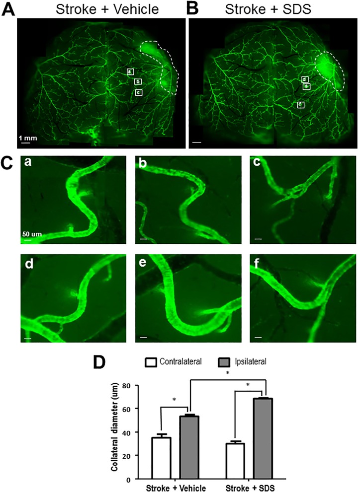
Fig. 5.
Enhanced collateral dilation by sodium danshensu.
The diameter of collaterals was measured by the imaging software ImageJ 21 days after stroke. (A, B) The arteries’ image of vehicle control mice (A) and SDS-treated mice (B). (C) The enlarged representative collaterals in vehicle control mice (a, b, c) and SDS-treated mice (d, e, f). (D) Mean diameter of collaterals in each group. Increased diameter of ipsilateral collaterals was shown 21 days after stroke and the dilation of ipsilateral collaterals was enhanced by SDS treatment. Six collaterals and six areas of each collateral were measured. The data are presented as mean ± SEM. * P < 0.05 compared with vehicle control group. SDS: sodium danshensu; SEM: standard error of the mean.

