Fig. 4.

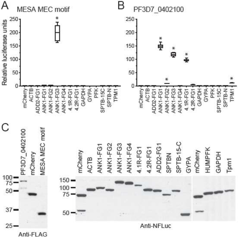
MEC motif proteins bind to erythrocyte ankyrin in the split-luciferase assay.
A. The MESA MEC motif binds to erythrocyte ankyrin. Proteins were in vitro translated in wheat germ extracts as fusions to N- and C-terminal fragments of firefly luciferase (N-FLuc and C-FLuc, respectively), mixed, and assayed for luciferase activity. Average relative luciferase activity (RLA) (± SEM) was calculated from duplicate readings from three independent replicates (six readings total) and was normalized to MESA-MEC motif plus N-FLuc-mCherry, which was set to 1. * indicates P value ≤ 0.001, as determined by one-way ANOVA with multiple comparisons to N-FLuc-mCherry using Graphpad Prism 6 software. ACTB, β-actin; ANK1, Ankyrin 1; 4.1R, Band 4.1; 4.2R, Band 4.2; GYPA, glycophorin A; GAPDH, glyceraldehyde phosphate dehydrogenase; SPTB-N, N-terminal fragment of β-spectrin; SPTB 15-C, β-spectrin fragment spanning spectrin repeat 15th to the C-terminus; ADD2, β-adducin; PFK, phosphofructokinase; TPM1, Tropomyosin 1.
B. PF3D7_0402100 binds to ANK1, 4.1R and TPM1 in the split-luciferase assay. Average RLA of C-FLuc-PF3D7_0402100 was calculated as above except that values were normalized to PF3D7_0402100 plus N-FLuc-mCherry. (* P ≤ 0.001).
C. Western blots showing the relative amounts of N-FLuc and C-FLuc fusion proteins used in panels A and B.
