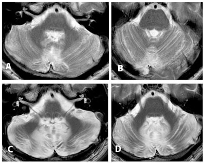
Figure.
T2-weighted brain MRI axial sections though the caudal and mid-pons following resection via vermis spitting approach of a choroid plexus papilloma. A, B: 2 years post-operatively, age 19. C, D: 15 years post-operatively, age 34. There is progressive volume loss, and signal hyperintensity in cerebellar white matter consistent with gliosis. The patient experienced cerebellar mutism, cerebellar cognitive affective syndrome, cerebellar motor syndrome, cranial neuropathies and corticospinal signs.

