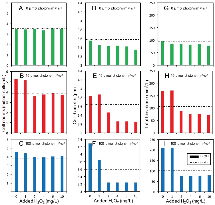
Figure 4.
Cell counts (A–C), cell diameter (D–F) and total biovolume (G–I) of Microcystis PCC 7806 at 24 h after addition of different H2O2 concentrations. Microcystis PCC 7806 was exposed to H2O2 at three different light intensities: (A,D,G) 0 µmol photons·m−2·s−1, (B,E,H) 15 µmol photons·m−2·s−1 and (C,F,I) 100 µmol photons·m−2·s−1. The bars represent the average of two independent biological replicates per treatment at 24 h after H2O2 addition. For comparison, horizontal, dashed lines indicate the cell counts (A–C), cell diameter (D–F) and total biovolume (G–I) prior to H2O2 addition (t = 0 h).

