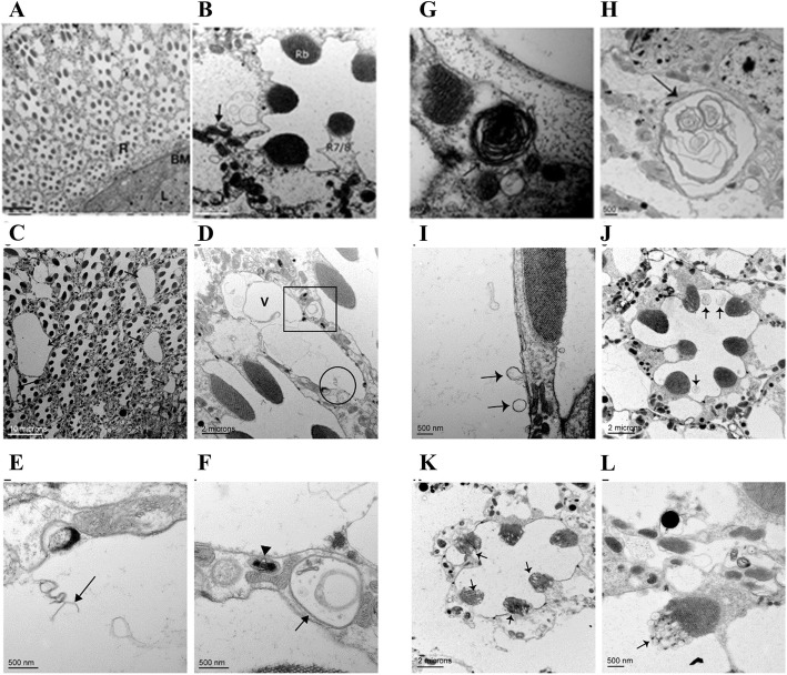
Fig. 3.
EM analysis of retinal degeneration in the Adar5G1 mutant. a The ommatidia of w1118 at 25 days. Each ommatidium comprises seven photoreceptor cells surrounded by and separated from neighboring ommatidia by thin pigment cells containing red pigment granules. b An ommatidium of 25-day-old w1118 at higher resolution. The photoreceptor cells with light-detecting rhabdomeres (Rb) appear normal. The R7/R8 photoreceptor is indicated. Organelles such as mitochondria are identifiable (arrow). c Retina of the Adar5G1 mutant at 25 days showing pigment cells with large vacuoles between ommatidia (arrows). d Higher resolution image of a single ommatidium in 25-day-old Adar5G1 with vacuole (V) between photoreceptor cells of two ommatidia. e Magnification of area within the circle in d. Interrupted membrane (arrow) was observed inside the vacuole. f Magnification of area within the square in d. Membrane-bounded vesicles (arrows) in the photoreceptors contain cellular components in an autophagosome-like structure surrounded by two or more membrane layers. g, h Multilamellar membrane structures (arrows) in a photoreceptor cell and within a glial cell close to the basement membrane between the retina and the lamina in Adar5G1. i Single membrane-bounded vesicles pinching off from the photoreceptor (arrows) in early stages of photoreceptor degeneration in Adar5G1. j Larger multilamellar membrane structures budding off from the extracellular membrane of photoreceptor cells into the ommatidial cavity (arrows) at more advanced stages of degeneration in Adar5G1. k Extensive loss of pigment cells separating ommatidia in advanced stages of neurodegeneration in Adar5G1. Photoreceptor cell cytoplasm and extracellular membrane are abnormal, and vesicles bud from the rhabdomeres (arrows). l Abnormal exocytosis from the rhabdomere in late stages. The extracellular membrane of the photoreceptor is not well defined

