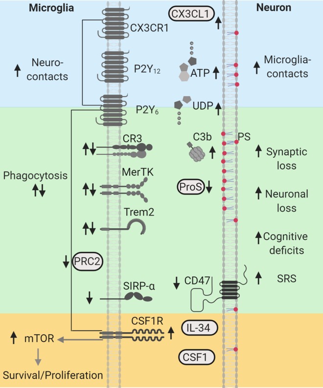
Figure 1.
Phagocytic signaling molecules altered in human and experimental epilepsy. “Find-me” signals CX3CL1/CX3CR1, ATP/P2Y12, and UDP/P2Y6, shown in blue, are associated with increased neuroimmune interactions during seizures. Microglia clearance/phagocytic activity controlled by PRC2 and mediated by “eat-me” signals PS (red), C3b/CR3, ProS/MerTK, and Trem2, shown in green, are associated with neuronal/synapse loss, cognitive deficits, and spontaneous recurrent seizures (SRS). “Don’t-eat-me” signals, CD47 and SIRP-α, shown in green, are reduced in human epilepsy. CSF1R-mTOR signaling activated by CSF1/interleuklin-34 (IL34), shown in yellow, regulate microglial survival, proliferation, and phagocytic microglial properties, and are associated with synaptic loss, cognitive decline, and SRS. Arrows indicate the direction of the changes reported in human and experimental models. This diagram was created with Biorender.com. CR indicates complement receptor; CSF1R, colony stimulating factor 1 receptor; MerTK, Mer Tyrosine Kinase; mTOR, mechanistic target of rapamycin; P2Y12, purinergic receptors; ProS, Protein S; PRC2, Polycomb repressive complex 2; PS, phosphatidylserine; SIRP-α, signal regulatory protein α; SRS, spontaneous recurrent seizures; Trem2, triggering receptor expressed in myeloid cells 2.

