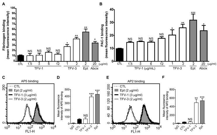
Figure 3.
Effect of TFV-1 and TFV-3 on the conformational change of αIIbβ3, ligand-induced binding sites (LIBS) exposure, and intrinsic monoclonal antibody recruitment. (A,B) Eptifibatide (Ept), abciximab, or various concentrations of TFV-1 or TFV-3 were added to human PS and then the platelets were fixed with 1% paraformaldehyde. After quenching the paraformaldehyde with glycine and washing, fluorescent fibrinogen ((A), 200 μg/mL) or PAC-1 ((B), 10 μg/mL) was added for 30 min at 37 °C. After washing twice, bound fluorescent fibrinogen or PAC-1 was detected by flow cytometry. The data shown are the mean fluorescence intensity of platelets in the presence of each compound. (mean ± SEM, error bars, n ≥ 5, *p < 0.05, **p < 0.01 and ***p < 0.001 compared with the control group by paired Newman–Keuls test; NS, non-significance) (C–F) Washed human platelets were incubated with PBS (Ctl), Eptifibatide (Ept, 2 μg/mL), TFV-1 (3 μg/mL) or TFV-3 (2 μg/mL), and then probed with 20 μg/mL mAb AP5 (C,D) or AP2 (E,F) raised against the ligand-induced binding site and the intrinsic antibody binding site of αIIbβ3, respectively. Levels of AP5 and AP2 binding to αIIbβ3 were analyzed by flow cytometry with fluorescein isothiocyanate-conjugated anti-IgG mAb as a secondary antibody. (mean ± SEM, error bars, n ≥ 6, **p < 0.01 and *** p < 0.001 compared with the control group by paired Newman–Keuls test; NS, non-significance).

