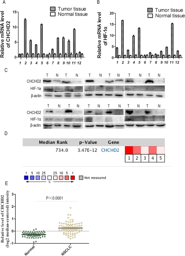
Fig. 1.
CHCHD2 and HIF-1α overexpressed in NSCLC tissue. a Expression of CHCHD2 mRNA was higher in tumor tissue than that in paired normal tissue. b Expression of HIF-1α mRNA was higher in tumor tissue than that in normal tissue. c Expression of CHCHD2 and HIF-1α protein were higher in tumor tissues (T) than those in pair normal tissues (N), and β-actin was used as an internal control. d A meta-analysis of CHCHD2 mRNA expression from Oncomine databases where colored squares indicate comparative analysis of CHCHD2 expression in specific data subsets. (1) LCC vs. Normal, Hou Lung [19], (2) ADC vs. Normal, Hou Lung [19], (3) SCC vs. Normal, Hou Lung [19], (4) ADC vs. Normal, Okayama Lung [20], (5) ADC vs. Normal, Su Lung [21], e Expression of CHCHD2 mRNA was higher in tumor tissue than that in normal tissue

