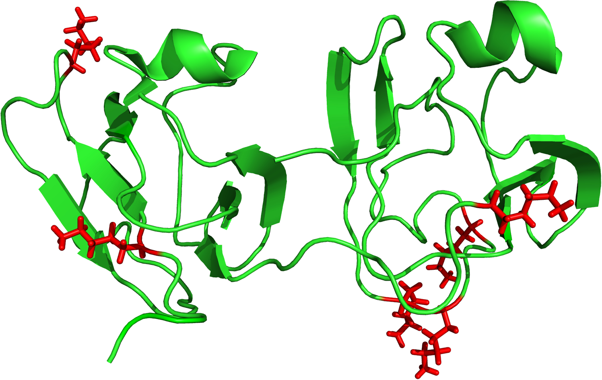
Fig. 2.

The structure of human gamma S crystallin derived from NMR data, where residue Gly18 was mutated to Val (PDBe 2M3U). Four of the five sites of attachment of DHAGly to Lys that are highlighted (red), occur within unstructured regions with the other site located at the boundary of a structured and unstructured region
