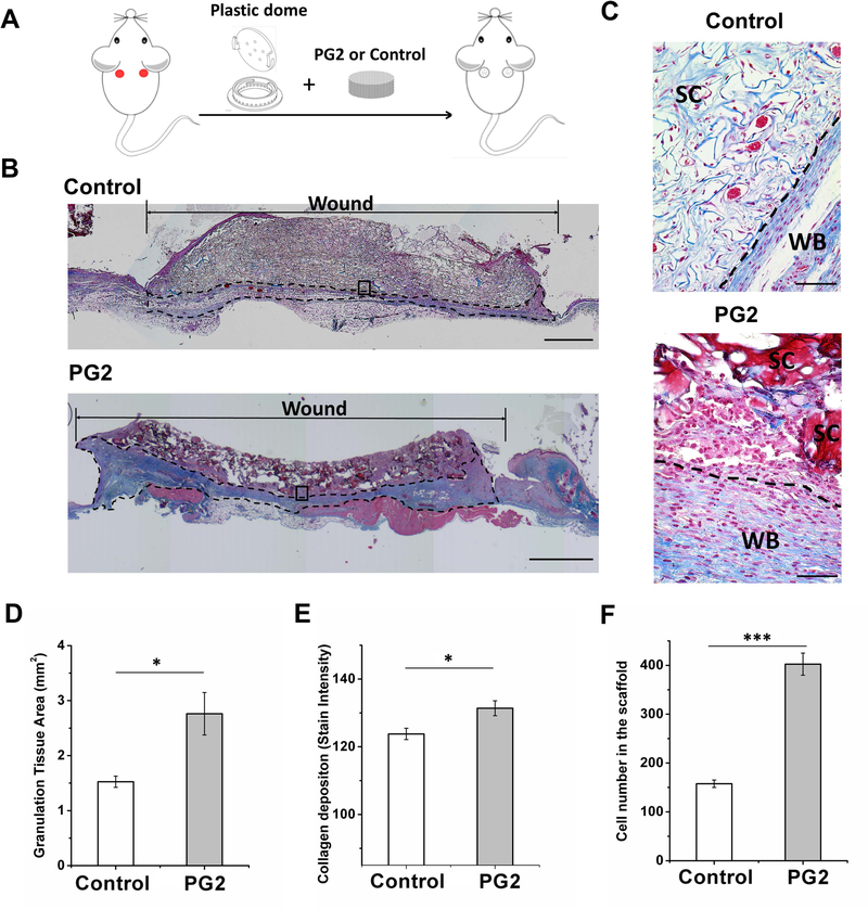
Figure 4.
PG2 enhance grannnulation tissue formation with higher cellularity and more collagen deposition. Scheme of the mouse model (A); representative Masson’s trichrome stained images to show a complete view of wounds treated with PG2 and Control by day 20, the dotted line marks collagen formation (B), scale bar = 1 mm; histological sections at 40X magnification showed scaffold fragments of PG2 and an integrated layer of Control (C), scale bar = 50 μm, SC, the scaffold or the scaffold fragment; WB, wound bed and collagen fiber deposition; quantification of granulation tissue area (D), collagen depositon (E) and infiltrated cell number (F) to demonstrate rapid, efficient and functional formation of neo-tissue. *p < 0.05, **p < 0.01, ***p < 0.001.

