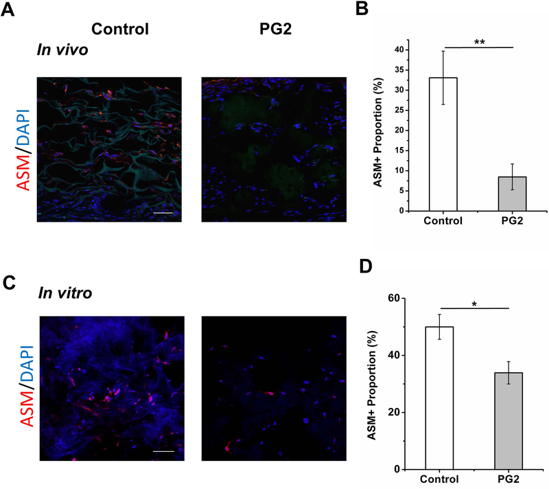
Figure 6.
The PG2 scaffold enhances less myofibroblastic phenotype. Immunofluorescent sections of wounds dressed with PG2 and Control on day 20 (A) and HDFs cultured in PG2 and Control in vitro 48h after TGF-β1 induction, stained with ASM (myofibroblast), to demonstrate myofibroblast differentiation in different scaffolds(C), scale bar = 100 μm; quantification results of ASM+ve cell proportion on wounds with different treatments in vivo (B) and in different scaffolds in vitro (D).

