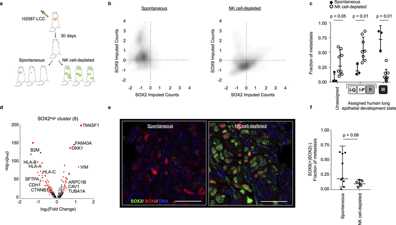
Figure 6. NK cell-dependent pruning limits the phenotypic expansion of metastasis-initiating cells.
a, in vivo NK cell perturbation assay in mice harboring latent metastasis-initiating cells. b, 2D cell density plot of z-normalized SOX2 and SOX9 imputed expression in H2087-LCC cells isolated from macrometastases +/− NK cell depletion, as determined by scRNA-seq. c, Fraction of type I/II and type III cells detected in spontaneous versus NK cell-depleted macrometastases (3 spontaneous macrometastases harvested from n = 3 independent mice and 8 NK cell-depleted macrometastases harvested from n = 5 independent mice; center line, geometric mean; whiskers, geometric s.d.; points, all measured data; two-sided Mann-Whitney rank test). Cell types are assigned by significant correlation with patient tumour states (Pearson R > 0.20 and two-sided p < 0.05 to test for non-correlation; as in Fig. 4f). d, Top DEG for NK cell-depleted cluster with highest SOX2 expression (Phenograph cluster 8, n = 322 cells, see Extended Data Fig. 10e) compared to all other cells, computed using MAST44. DEGs are red, with diameter proportional to −log10(padj) for genes with fold change > 1.5 and padj < 0.05. e, SOX2 and SOX9 immunofluorescence in a representative spontaneous and NK cell-depleted macrometastasis (n = 15 macrometastases evaluated, nuclear SOX9 expression summarized in f). Scale bars, 50 μm. f, Nuclear SOX2 and SOX9 single-positive, double-positive, and negative cell fractions quantified per macrometastatic lesion (n = 11,376 single cells quantified, fraction of metastases reported across n=15 lesions including lung, bone, kidney, and soft connective tissues harvested from 7 mice). 5 representative 20X frames were evaluated per lesion. SOX9 single-positive cells were enriched in spontaneous as compared to NK-cell-depleted macrometastases (n = 15 independent macrometastases, p = 0.06, one-sided Mann-Whitney rank test); abundance of other cell types was not significantly altered (data not shown). Center line, geometric mean; whiskers, geometric s.d.; points, all measure data.

