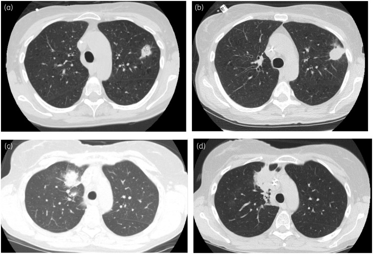
Figure 1.
(a) CT scan of Patient 1 obtained on day +10 after transplant immediately prior to initiation of voriconazole, demonstrating left upper lobe pulmonary nodule. (b) CT scan of Patient 1, obtained on day +35 after transplant, demonstrating interval enlargement and increase in density of the left upper lobe nodule. (c) CT scan of Patient 2, obtained on day +11 after transplant, immediately prior to initiation of voriconazole. (d) CT scan of Patient 2, obtained on day +27, demonstrating worsening right upper lobe consolidation.

