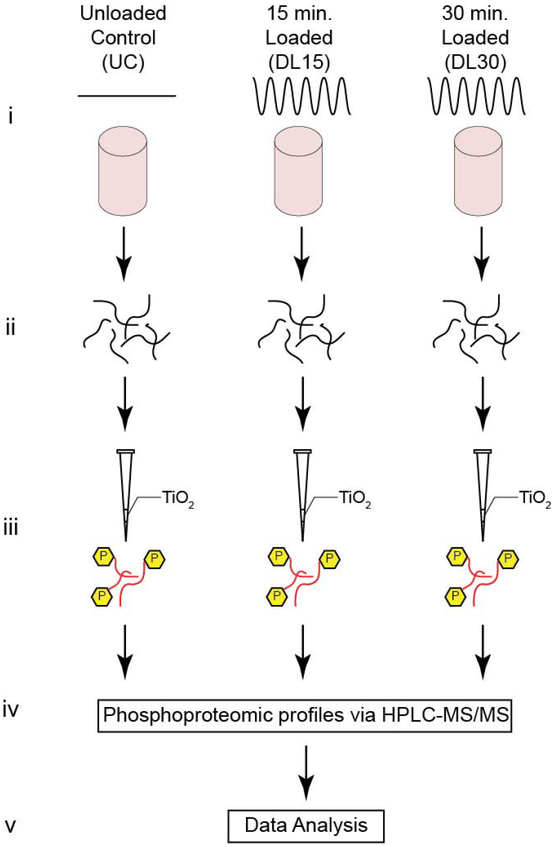
Figure 1. Experimental Design.
(A) Schematic for both untargeted experimental methods. (i) Primary human OA chondrocytes are encapsulated in physiologically stiff agarose (4.5% agarose, stiffness ~35 kPa), cultured for 72 hours, and then dynamically compressed in tissue culture for 0, 15, or 30 minutes (Control, DL15, or DL30) at 1.1 Hz. (ii) Proteins are extracted by flash freezing the samples, pulverizing, and lysing the cells followed by overnight enzymatic digestion. (iii) Samples are enriched for phosphopeptides using TiO2 enrichment, (iv) phosphoproteomic profiles identified via HPLC-MS/MS, and (v) the untargeted data analyzed.

