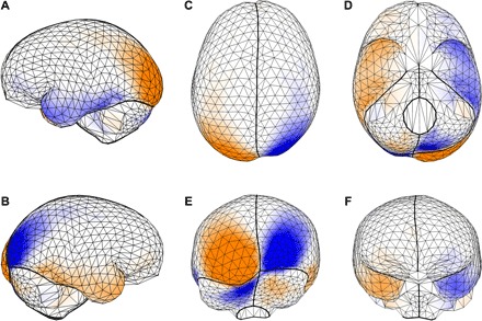
Fig. 3.

Shared directional shape asymmetry pattern. PC 1 of endocranial shape asymmetry in Fig. 2B is shown as a triangulated surface mesh of the 935 (semi)landmarks in (A) left, (B) right, (C) superior, (D) inferior, (E) occipital, and (F) frontal views. The deformation from a symmetric endocranial shape represents the spatial pattern of shape asymmetry; orange surfaces have larger areas as compared with the other side, and blue surfaces have smaller areas. See also movie S1.
