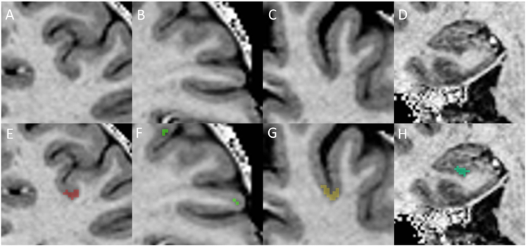
Figure 3: Examples of cortical lesion subtypes and cortical lesion masking.
Cortical lesions were identified as hypointensities on T1w MP2RAGE images with associated hyperintensity on MPFLAIR (not shown). The upper panel is unmarked, and the lower panel shows the lesion area inpainted as a colored mask. A/E = Leukocortical, B/F = Intracortical, C/G = Subpial, D/H = Hippocampal.

