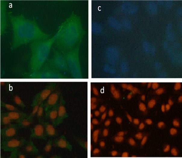
Figure 6.
Immunofluorescence Staining of Pari-ICR Cell Line. Expression of Ezrin and Vimentin in pari-ICR was investigated by imunofluorescence assay. (a) Immunofluorescence staining with anti-Ezrin antibody, (b) Immunofluorescence staining with anti-Vimentin antibody, (c,d) Negative controls. Nucleus is stained by DAPI in figures (a) & (c), and by PE in figures (b) & (d)

