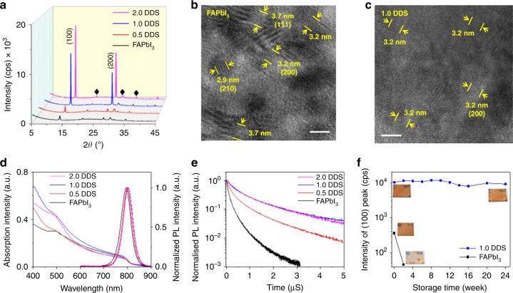
Fig. 2. Properties of PMC thin films.
a XRD patterns of perovskite films; diffraction peaks from the ITO substrate are marked as ♦. Cross-sectional HRTEM images of (b) the FAPbI3 control film and (c) the 1.0 DDS film. The measured lattice spacings (10 fringes for each pair of markers) match well with the cubic α-phase FAPbI3 structure. The scale bars for (b) and (c) are 5 nm. d Absorption and PL spectra and e time-correlated single-photon counting spectra (recorded at a fluence of 0.13 μJ cm−2) of perovskite films. f XRD intensity of the (100) diffraction peak of perovskite films (inset: photographs of the fresh and aged perovskite films).

