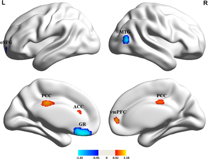
Fig. 3.
Cortical thickness alterations in medication-free patients with major depressive disorder compared with healthy controls. Regions of increased (warm color) and decreased (cool color) cortical thickness in medication-free patients with MDD than HCs in the pooled meta-analysis. ACC, anterior cingulate cortex; GR, gyrus rectus; L, left; oSFG, orbital segment of the superior frontal gyrus; PCC, posterior cingulate cortex; vmPFC, ventromedial prefrontal cortex; MTG; middle temporal gyrus; R, right

