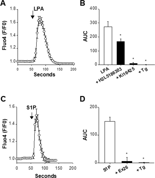
Figure 2.
Presence of functional LPA- and S1P-sensitive receptors. The presence of functional LPA and S1P receptors was verified with the fluorescent Ca2+ probe Fluo-4. In these experiments, cells were maintained in a nominally Ca2+ free saline. Panels A and C show somatic Fluo-4 signals (F/F0) as a function of time in response to 10 µM LPA (n = 10) (panel A) and 10 µM S1P (n = 9) (panel C). Panel B shows the LPA-induced Ca2+ rises measured as area under the curve (AUC) in the absence (white bar, n = 10) or presence of H2L5186303 (10 µM, n = 5), Ki16425 (10 µM, n = 7), or after the application of thapsigargin (Tg, 200 nM, n = 5). *p < 0.05 vs LPA, one-way ANOVA followed by a Bonferroni’s post hoc test. Panel D shows the Fluo-4 responses (measured as area under the curve, AUC) induced by S1P alone (10 µM, n = 9), S1P + Ex26 (1 µM, n = 7), and S1P applied after thapsigargin (Tg, 200 nM, n = 5), with *p < 0.05 vs S1P, one-way ANOVA followed by a Bonferroni’s post hoc test. Antagonists of LPA and S1P receptors were added 4–7 min before time 0 and remained present throughout the recordings. LPA and S1P can stimulate store-released Ca2+. Pre-depleting the ER Ca2+ with Tg prevents any response to LPA or S1P.

