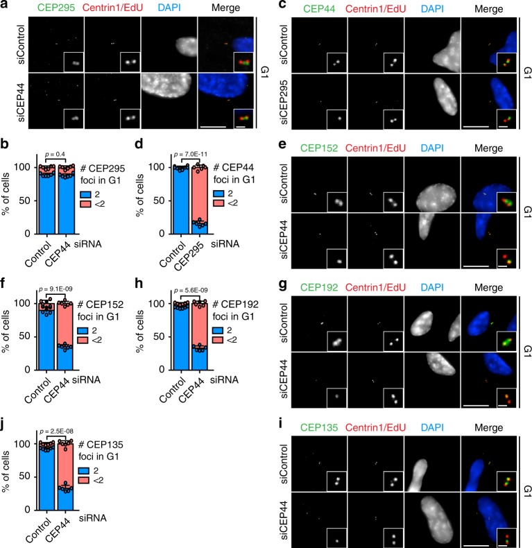
Fig. 2. CEP44, downstream of CEP295, influences the conversion mechanism.
a CEP295 did not delocalise from the centrioles in G1 cells (Cenp-F in Supplementary Fig. 3a) upon CEP44 siRNA depletion (10.9 ± 2.3% of cells with <2 CEP295 foci in siControl cells vs. 12.1 ± 2.5% in siCEP44 sample). b Quantification of a. c CEP44 strongly delocalised from the daughter centrosome in the absence of CEP295. G1 cells (Cenp-F negative in Supplementary Fig. 3c) were analysed. d Quantification of c. In siCEP295 84.0 ± 3.7% of the cells had <2 CEP44 foci. e CEP44-less daughter centrosomes lacked CEP152 (f, 64.9 ± 3.2% of G1 cells vs. 10.1 ± 5.1% in control; Cenp-F in Supplementary Fig. 3i, left column), g CEP192 (h, 67.1 ± 3.6% of G1 cells; Cenp-F in Supplementary Fig. 3i, middle column) and i CEP135 (j, 66.9 ± 4.6% of G1 cells; Cenp-F in Supplementary Fig. 3i, right column). (a, c, e, g, i, scale bars: 10 μm, magnification scale bars: 1 μm; b, d, f, h and j data are presented as mean ± s.d., all statistics were derived from two-tail unpaired t-test analysis of n = 6 biologically independent experiments and source data are provided as a Source Data file).

