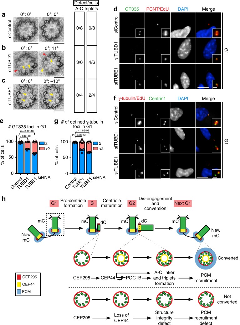
Fig. 7. Structural integrity of the centrioles ensures their conversion to centrosomes.
a–c EM images of turned and tilted sections of proximal region of daughter centrosomes in G1. The two values on the top of the image give the turning and tilting degree of the sample, respectively. Red arrowheads indicate open structural defects (A–C linker absent), while yellow ones highlight defects of triplets. The side table (right) shows quantification of the number of cells showing these defects. While in a G1 siControl cells had no structural defects, in b siTUBD1 and c siTUBE1 G1 dCs showed defects in triplet formation (yellow arrow). b siTUBD1 G1 cell with defect in the A–C linker formation on top of the triplet formation of dCs and lack of triplet MTs (red arrow). d Depletion of TUBD1 and TUBE1 proteins also generated reduced glutamylation of the centrioles as judged by GT335 staining (36.7 ± 2.2% of G1 cells, Cenp-F in Supplementary Fig. 12a) with <2 GT335 foci in siTUBE1 sample; 23.5 ± 2.0% for siTUBD1). e Quantification of d. f In absence TUBE1 or TUBD1 many centrioles in G1 cells (Cenp-F in Supplementary Fig. 12b) did not efficiently acquire PCM (<2 γ-tubulin defined foci) to convert to centrosomes. g Quantification of f. 33.1 ± 1.3% of cells with <2 γ-tubulin foci in G1 for siTUBE1; 26.2 ± 4.1% for siTUBD1. (a–c, scale bars: 100 nm; d, f scale bars: 10 μm, magnification scale bars: 1 μm; e, g data are presented as mean ± s.d., all statistics were derived from two-tail unpaired t-test analysis of n = 6 biologically independent experiments and source data are provided as a Source Data file). h Model of the dependency of CCC mechanism on the correct centriolar wall maturation. See Discussion for description.

