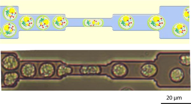
Figure 1.
Mechanical compression of C. reinhardtii cells in the microfluidic system. Upper image: schematic drawing of the mechanical compression process; the electroporated cells, enriched in compounds of interest (lipids, protein, pigments…), are compressed by flowing through the microfluidic restrictions (height/width: 5 µm) leading to a higher permeabilisation of the cell. Lower image: observation of the mechanical compression of C. reinhardtii cells under bright field microscopy.

