Figure 3.
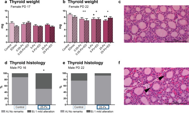
Thyroid gland weight and histology PD 16/17 and 22 after developmental exposure to PFHxS. (a,b) Female pup thyroid gland weight at PD 17 and PD 22. Data shown as mean + SEM. n = 11–16. (d,e) Thyroid histopathology on male pup thyroid glands PD 16 and PD 22. Bars represent percentage of animals receiving indicated score. control n = 16–17 and 25-Px n = 13–14. (c,f) Representative images of thyroid tissue from a control male pup (c) receiving a score of A (no remarks) and a male pup from the 25-Px group (f) receiving a score of B (1 mild alteration, potentially within natural variation) for altered cellularity (arrowheads). * p < 0.05 compared to control, **p < 0.01 compared to control, +p < 0.05 for full model comparison of indicated dose of PFHxS compared to no PFHxS exposure in the control and EDmix group. ++p < 0.01 for full model comparison of indicated dose of PFHxS compared to no PFHxS exposure in the control and EDmix group. ED: EDmix. Px: PFHxS. PD: postnatal day.

