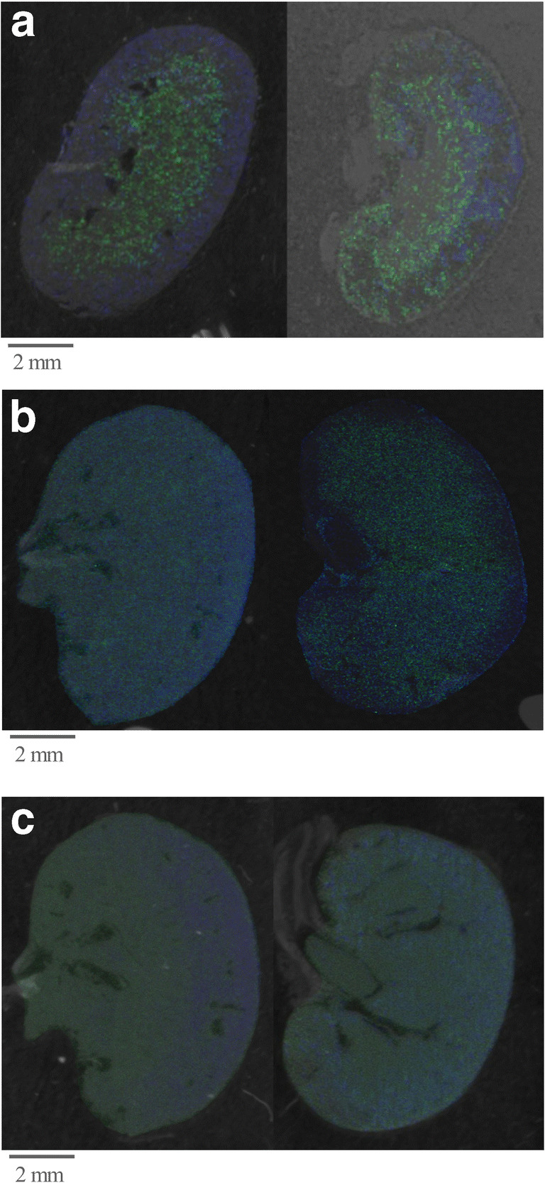
Fig. 5.

MALDI MSI images of fresh-frozen tissues (left image) compared with FFPE tissue samples (right) using the optimized SOP. Shown are MALDI MSI images of a mouse kidney using the optimized SOP and overlaid on the tissue scan. 718 (m/z) green, 958 (m/z) blue (a, b accumulated by using an Ultraflex mass spectrometer; c accumulated by using Rapiflex mass spectrometer (both from Bruker Daltonics, Bremen, Germany)). a Partial resolution 50 μm. b Partial resolution 25 μm. c Partial resolution 10 μm
