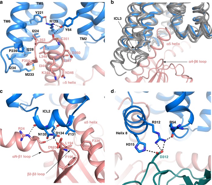
Fig. 6. FPR2 and Gi interface.
a Interactions between α5N of Gαi (salmon) and the receptor (blue) in the cavity at the cytoplasmic region of FPR2. b Comparison of the cytoplasmic regions of TM5 and TM6 and ICL3 in FPR2 (blue) and in μOR (PDB ID 6DDE), A1AR (PDB ID 6D9H), CB1 (PDB ID 6N4B) and rhodopsin (PDB ID 6CMO) (all dark gray). Gαi in the structure of FPR2-Gi is shown in salmon. c Interactions between the ICL2 of FPR2 and Gαi. d Interactions between FPR2 and Gβ.

