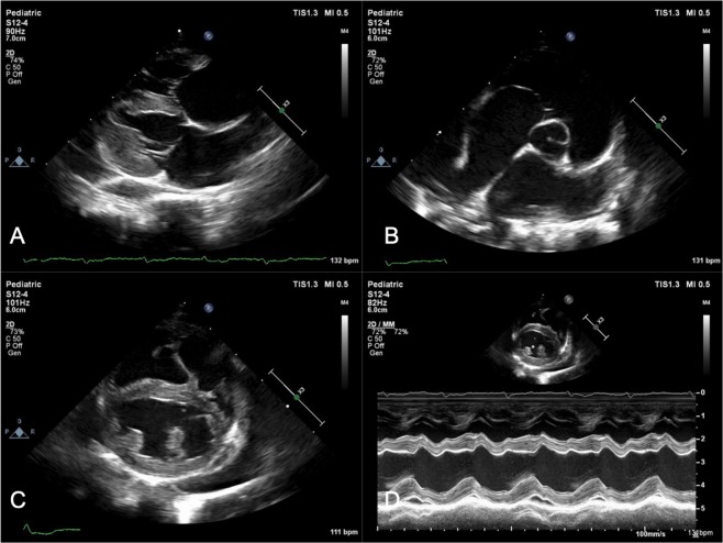
Figure 1.
Echocardiographic images from a representative feline patient. (A) Demonstrates a right parasternal long-axis 4-chamber view and (B), a right parasternal short-axis basilar view both depicting severe biatrial enlargement with scant pericardial effusion. Two-dimensional imaging (C) and M-mode (D) from the same feline patient at the level of the left ventricular papillary muscles. There is equivocal myocardial thickening, left ventricular dilation, systolic dysfunction (Fractional shortening = 27%), and decreased septal wall motion on M-mode. Necropsy findings in this patient demonstrated dissecting fibrosis with multifocal myonecrosis and hypertrophy.

