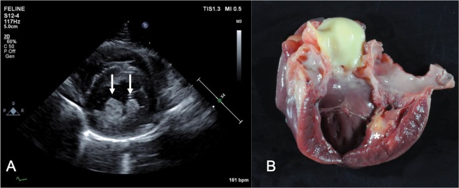
Figure 4.
Echocardiographic (A) and gross pathology (B) images from one feline patient. The echocardiographic image depicts the right parasternal short-axis view at the level of the papillary muscles. The papillary muscles are prominent and hyperechoic (arrows). Gross pathology image demonstrated a pale multi-nodular focus within the papillary muscle (*) and extending through the left ventricular free wall. Histopathology of this region demonstrated severe myocarditis with intralesional thrombi and coccoid bacteria. Photo courtesy of Dr. Melissa Roy.

