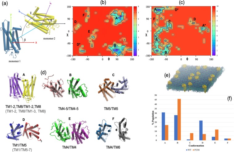
Figure 5.
(a) Angles used to describe the orientation of the monomers within the receptor dimers. Explicitly, β describes the position of monomer 2 with respect to monomer 1, χ is the angle that designates the contact orientation between monomers 1 and 2, and ϕ is the rotation of monomer 2 about its z-axis. The sampled (b) WT and (c) P23H homodimer configurations from the CG simulations. (d) A cartoon representation of the dimer configurations as labeled in (b) and (c). The labeling in gray refers to the slightly different dimer structures in the P23H self-assembly homodimer system when compared with the WT rhodopsin self-assembly system. (e) A typical early timescale arrangement of the self-assembly simulations comprising 16 receptors. (f) Relative populations of the dimer configurations found in the respective CGMD self-assembly simulations.

