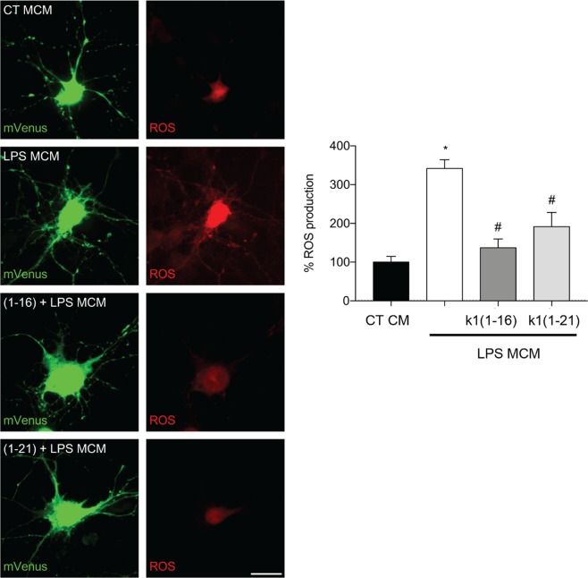
Figure 7.
Ocellatin-K1(1–16) and Ocellatin-K1(1–21) protect hippocampal neurons from oxidative stress induced by LPS-treated-microglial conditioned media. Representative confocal images and quantification of ROS production in hippocampal neurons incubated with conditioned medium from microglia subjected to LPS-induced and 100 μM Ocellatin-K1(1–16) or Ocellatin-K1(1–21). Images show neurons expressing mVenus (green) and the Hyper Red ROS biosensor (red). The results are expressed as mean ± SEM calculated from 3 different cultures. *p < 0.001 vs. LPS group; #p < 0.001 vs. CT employing one-way ANOVA with the Bonferroni post-test. Abbreviations: CT: control; LPS: lipopolysaccharide; MCM: microglia conditioned medium.

