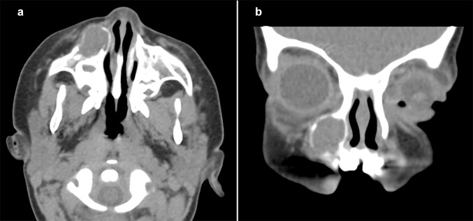
Fig. 1.
Axial (a) and coronal (b) views on CT scans demonstrate a well-circumscribed, expansile, hypodense soft tissue mass with a peripheral hyperdense circumference in the right maxilla, anterior to the right maxillary sinus. a Encroachment of the anterior wall of the right maxillary sinus and right lateral nasal wall without invasion of nearby structures. b The lesion extends from the inferior aspect of the orbital rim superiorly to the maxillary bone inferiorly

