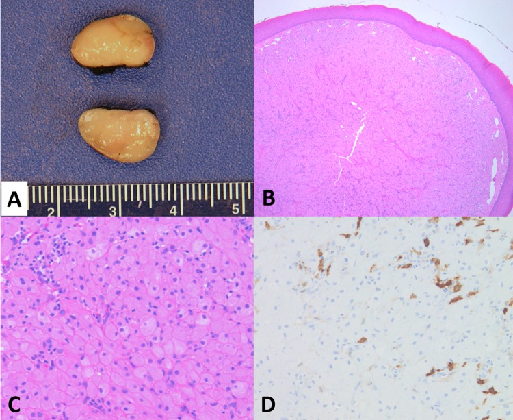
Fig. 1.
a Gross examination of CGCE shows a homogeneous, tan-yellow, and smooth cut surface. b Low power view of the lesion shows a lobulated mass with overlying, thin squamous epithelium (H&E, × 4). c The mass is characterized by proliferation of polygonal cells with eosinophilic, granular cytoplasm and eccentric, benign-appearing nuclei. Scattered interstitial and inflammatory cells are noted in the upper left of the image (H&E, × 10). d Unlike granular cell tumor in adults, lesional cells of CGCE are negative for S-100 immunostain; the S-100-positive cells in the image represent interstitial cells (× 10)

