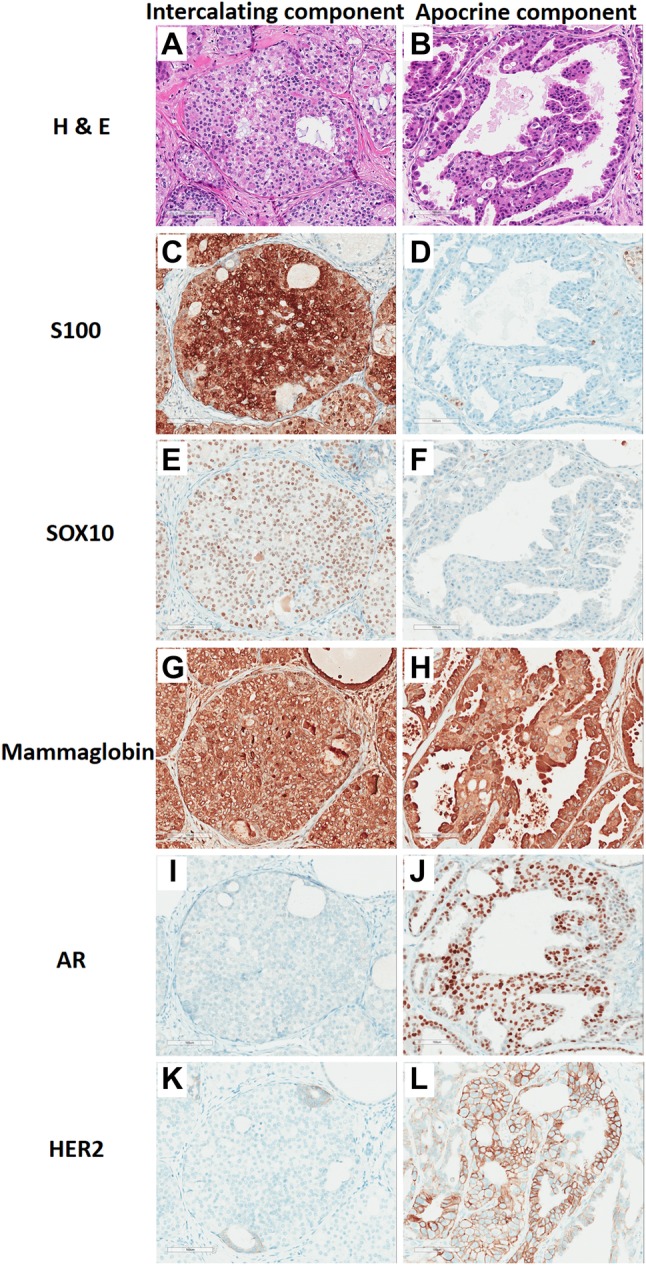
Fig. 4.

Surgical resection comparative immunohistochemistry. The intercalated duct component was monomorphic and bland with a nested clear cell pattern (a), and was strongly positive for S100 (c), SOX10 (e), and mammaglobin (g), and negative for AR (i) and HER2 (k). The apocrine component had larger nuclei, variable nucleoli, and had abundant eosinophilic cytoplasm (b), and not express S100 (d) and SOX10 (f), but strongly expressed mammaglobin (h) and AR (j) and partially expressed membranous HER2 (l). (20 ×, Bar = 50 µm)
