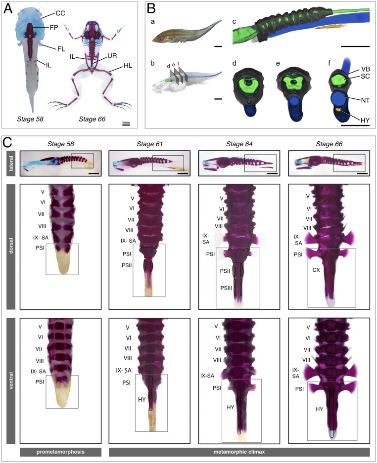
Fig. 1.
Bone and cartilage formation of the urostyle. (A) Dramatic skeletal remodeling at metamorphic climax in X. tropicalis visualized through cartilage and bone staining, using Alcian blue and alizarin red, respectively. Cartilage is depicted in blue; bone is depicted in red. The larval chondrocranium remodels and forms new cranial bones. The urostyle forms during metamorphic climax and lies between the two ilia. (B) CT-scanned tadpole of X. tropicalis, NF stage 61 (metamorphic climax, day 2), highlighting axial skeleton formation at the rostral end of the tadpole body. The coccyx and the hypochord form midlength of the body, and the tail resorbs completely during metamorphosis. (a) Photograph of a live NF-61 tadpole; (b) CT-scanned tadpole after volume rendering; (c–f) Segmented tadpole highlighting the spinal cord, axial column, notochord, and hypochord. (C) Coccyx and hypochord formation at metamorphic climax in X. tropicalis. The coccyx is initiated as two ossification centers, which extend posteriorly and anteriorly. The hypochord forms ventral to the notochord and fuses with the coccyx at the end of metamorphic climax. Dorsal and ventral views are higher magnification images of the selected areas of the lateral view of each corresponding stage. The selected vertebrae are numbered from I to IX. CC, chondrocranium; CX, coccyx; FL, forelimb; FM, femur; FP, frontoparietal; HL, hind limb; HY, hypochord; IL, ilium; NT, notochord; PS, postsacral; SA, sacrum; SC, spinal cord; UR, urostyle; VB, vertebrae. (Scale bars: 5 mm.)

