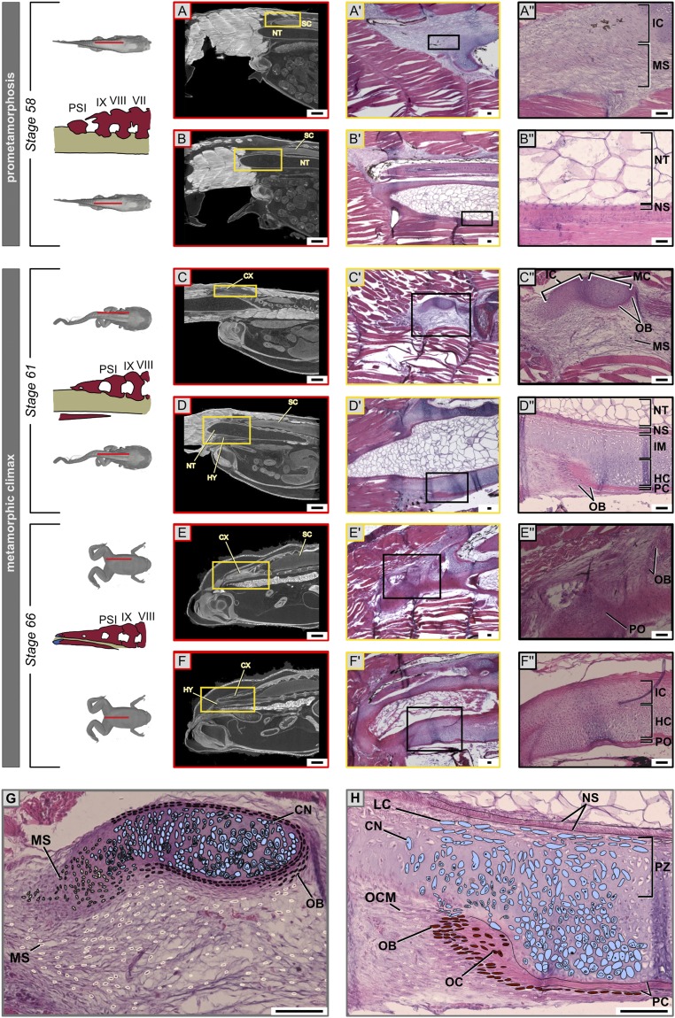
Fig. 2.
Chondrocyte–osteocyte differentiation during urostyle formation. (A–H) Hematoxylin (stains nuclei purple) and eosin (stains cytoplasm and extracellular matrix pink) staining of histological sections (sagittal, A′–F′) and orthoslices (sagittal, A–F) of CT scans of X. tropicalis at the site of urostyle formation. The Upper Row of each stage corresponds to a left parasagittal section. The Lower Row of each stage depicts a midsagittal section. Magnified cells in the last column (A″–F″) are cells of interest, which change during development. (A′, B′, A″, and B″) At stage 59, undifferentiated mesenchymal cells (scleroblasts) at the sites of the future coccyx aggregate to form mesenchymal condensations that form the rudimentary neural arches; osteo-chondro progenitors of the hypochord are present ventral to the notochordal sheath. (C′, D′, C″, and D″) Cartilaginous condensations are visible as immature chondrocytes and mature chondrocytes. Chondrocytes in the ossifying coccyx and hypochord; the perichondrium starts to form around the mature chondrocytes. (E′, F′, E″, and F″) The periosteum forms with the degeneration of the cartilaginous matrix, but, during hypochord ossification, hypertrophic chondrocytes degenerate, and some dedifferentiate into osteocytes. (G and H) Illustrations of ossification patterns of the coccyx and hypochord, highlighting the proliferating zone, growth zone, and ossifying zones for the two tissue types. CN, chondrocytes; CX, coccyx; ECM, extracellular matrix; HC, hypertrophic chondrocytes; HY, hypochord; IC, immature chondrocytes; IM, immature cartilage cells; LC, lacunae; MC, mature chondrocytes; MS, mesenchymal cells (scleroblasts); NS, notochordal sheath; NT, notochord; OB, osteoblasts; OC, osteocytes; OCM, osteo-chondro progenitors; PC, perichondrium; PO, periosteum; PZ, proliferating zone; SC, spinal cord. (Scale bars: 2 mm (A–F) and 100 μm.)

