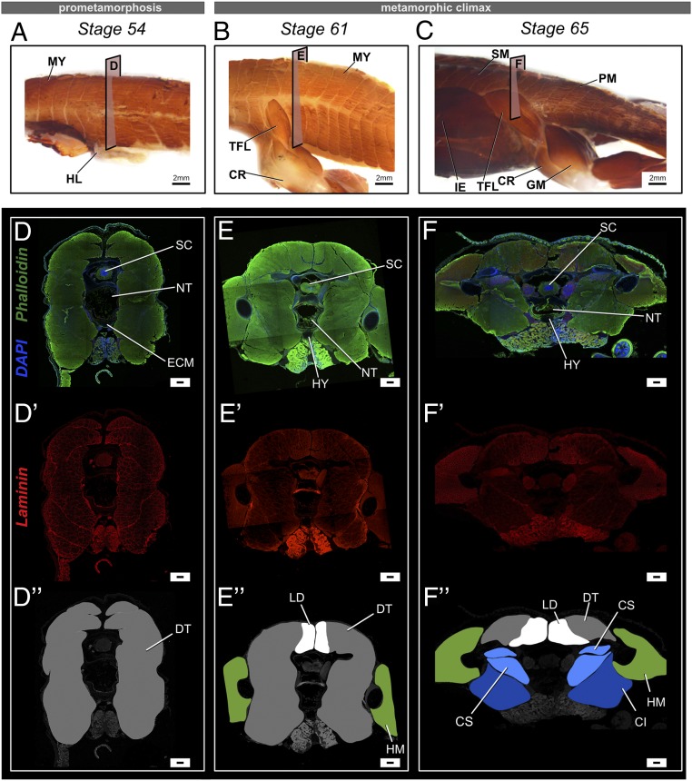
Fig. 3.
Changes in muscle composition near the urostyle before and after metamorphosis in X. tropicalis. (A–C) Lateral views of whole-mount immunostained specimens for skeletal muscle marker 12-101 at stages 54 (A), 61 (B), and 65 (C). Before metamorphic climax, primary muscles are undifferentiated and referred to as dorsal trunci (DT). During metamorphic climax, the myomeres undergo secondary myogenesis and form new muscle types, which differ in muscle fiber width and are attached to the newly forming skeletal structures. The coccygeo-iliacus originates from the lateral surface of the urostyle; the longissimus dorsi originates from the dorsal part of the urostyle and extends anteriorly; and the coccygeo-sacralis connects the sacrum and the coccyx. (D–F and D′–F′) Transverse cross-sections across the trunk myotome XII at prometamorphic and metamorphic stages, where phalloidin (green) stains the extracellular matrix, DAPI (blue) stains nuclei, and laminin (red) stains muscle fibers. (D, D′, and D″) Comparison of muscle fiber width shows that primary muscles have a constant width across the trunk body. (E′ and F′) Newly differentiating muscles (dorsal-most muscles) are smaller in fiber width. (D″–F″) Illustrations of the transverse cross-sections of the respective stages highlighting the different types of primary and secondary muscles. CI, coccygeo-iliacus; CR, cruralis; CS, coccygeo-sacralis; DT, dorsalis trunci; GM, gluteus magnus; HL, hind limb; HM, hind limb muscles; IE, iliacus externus; LD, longissimus dorsi; MY, myomeres; PM, primary muscles; SM, secondary muscles; TFL, tensor fasciae latae. (Scale bars: 2 mm (A–C) and 100 μm.)

