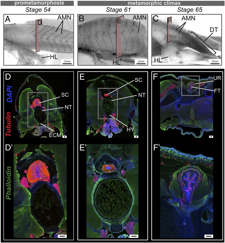
Fig. 4.
Changes in the spinal cord and innervation at the sites of urostyle formation during development. (A–C) Lateral views of whole-mount immunostained tadpoles using acetylated tubulin, at stages 54 (A), 61 (B), and 66 (C). Before tail regression is initiated, the axial myomeres possess axial motor neurons (AMNs), equally distributed, but AMNs degenerate with the regressing tail during metamorphic climax. (D–F) Transverse cross-sections across trunk myotome XII at prometamorphic and metamorphic stages, immunostained for acetylated tubulin (red), extracellular matrix (green), and nuclei (blue). (D′–F′) Magnified images of the spinal cord for each corresponding stage. (D, D′, E, and E′) The spinal cord is recognizable as gray matter (in the middle) and white matter (surrounding the gray matter) in transverse cross-sections. (F and F′) The spinal cord changes shape with the fusion of the coccyx and hypochord and is referred to as the filum terminale. AMN, axial motor neurons; DT, degenerating tail; ECM, extracellular matrix; FT, filum terminale; HL, hind limb; HY, hypochord; NT, notochord; SC, spinal cord; UR, urostyle. (Scale bars: 2 mm (A–F) and 100 μm.)

