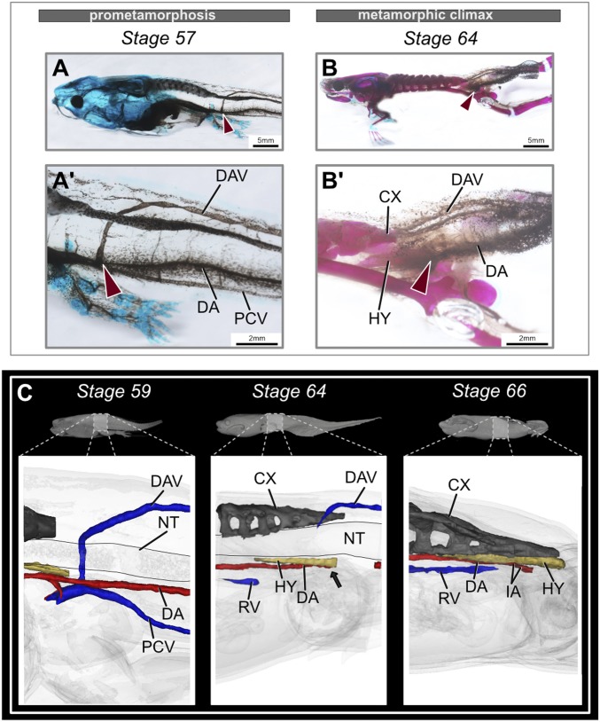
Fig. 5.
Rearrangement of the major blood vessels during metamorphosis. (A and B) Comparison of the dorsal aorta and posterior veins in the tadpole tail during metamorphosis, lateral view. Before metamorphosis, the DA and PCV are present ventral to the notochord, and the DAV is present dorsal to the spinal cord. The DAV then merges with the PCV. At the end of metamorphosis, stage-64 tadpoles lose the DA partially, along with PCV and DA. Red arrows point at the merging of DAV and PCV. (A′ and B′) Magnified images of the corresponding stage, highlighting the major blood vessels. (C) MicroCT-scanned tadpoles at prometamorphic and metamorphic climax stages. The black arrow points to the occlusion point of the DA at stage 64 (before the tail starts to regress). Stage 66 highlights the formation of new veins and rearrangement of the DA in the metamorphosed frog (refer to Datasets S1–S3 to observe this closely). CX, coccyx; DA, dorsal aorta; DAV, dorsolateral anastomosing vessel; HY, hypochord; IA, iliac artery; NT, notochord; PCV, posterior cardinal vein; RV, renal vein.

