Fig. 6.
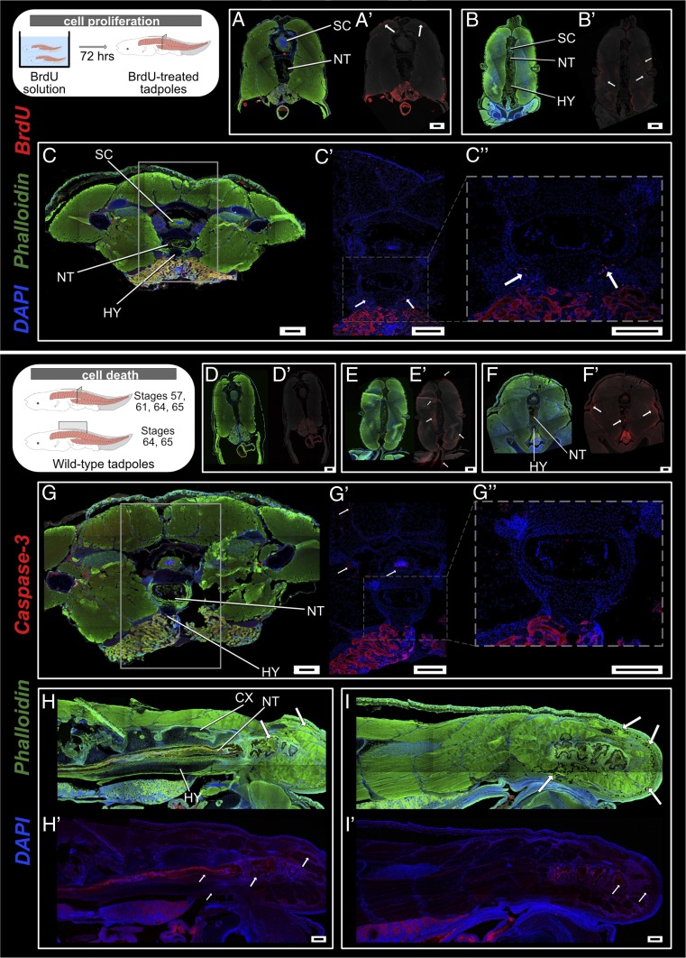
Cell death and cell proliferation at the site of urostyle formation. (A–C) Transverse sections across trunk myotome XII, immunostained for anti-BrdU (red) to observe cell proliferation at stages 58 (A and A′), 61 (B and B′), and 64 (C and C′). (A′–C′) Anti-BrdU signal has been overlaid on a grayscale background to highlight the proliferating cells. Muscle cells initiate proliferation prior to metamorphic climax, starting with the dorsal-most muscles (A′). With the initiation of metamorphosis, muscles along the lateral margins proliferate (B′). Lateral margins of the hypochordal rod demarcate the chondrocyte proliferating zone (C, C′, and C″). White arrows depict proliferating zones. (D–I) Transverse and sagittal cross-sections across the trunk myotomes, immunostained for anti-Caspase 3 (red) to observe cell apoptosis. There is no cell death at stage 57, before metamorphic climax (D and D′). With the initiation of metamorphic climax at stage 61 (E and E′), innermost muscles surrounding the notochord and ventral-most muscles undergo apoptosis (“suicide model”). Dorsal-most and lateral larval muscle fibers undergo cell death at stage 63 (F and F′). By stage 64, the coccyx and hypochord have reached the maximum length by extending up to the myotome, and the spinal cord is degenerated (G, G′, and G″). Sagittal sections at stages 64 (H and H′) and 65 (I and I′) depict how the muscle cells are degenerated by phagocytosis (“murder model”) with the reduction of the tail. CX, coccyx; HY, hypochord; NT, notochord; SC, spinal cord. (Scale bars: 100 μm.)

