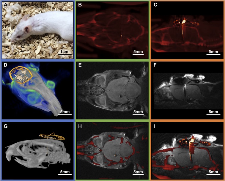
Fig. 4.
(A) Photograph of a mouse 20 d after implantation of a small photometry device. (B) Transverse CT reconstruction of the implanted device. (C) Sagittal CT reconstruction of a slice that illustrates the position of the implant. (D) Three-dimensional rendering that combines MRI (blue) and CT (gray and yellow marked device structures) results. (E) Transverse MRI reconstruction of the device illustrating imaging capability around the target area. (F) Sagittal MRI reconstruction of the device illustrating minimal distortion of the brain. (G) Postprocessed sagittal CT scan with false color device structures (yellow). (H) Transverse MRI and CT overlay of the device, indicating successful registration and low-dimensional image distortion in both MRI and CT. (I) Coregistered sagittal MRI and CT overlay.

