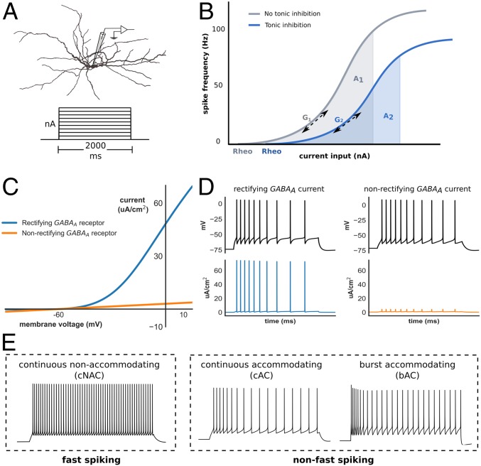
Fig. 1.
Interneuron modeling and measurement of gain. (A) Interneuron model morphology and step-current protocol used to obtain the I–F relationship. (B) The I–F relationship was obtained with (blue) and without (gray) tonic inhibition, and the normalized change in gain (Δ gain) defined as either the change in AUC (from A1 to A2) across a fixed input range, or the change in gradient (G1 to G2) at a spike frequency of 20 Hz (Methods; rheo denotes rheobase). (C) I–V relationship of rectifying (blue) and nonrectifying (orange) extrasynaptic GABAA receptors. (D) Rectifying extrasynaptic GABAA receptors allow greater outward (hyperpolarizing) current to be passed at transmembrane voltages above approximately −50 mV, such as during AP generation. (E) Time–voltage traces of three models optimized to exhibit different interneuron E-type classified according to either fast-spiking vs. non–fast-spiking categories, or Petilla E-type. Note: by convention, hyperpolarizing transmembrane current is positive.

