Fig. 3.
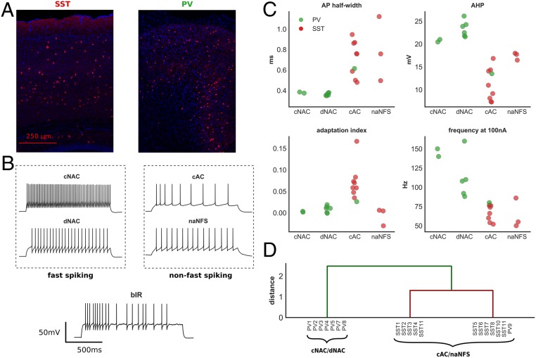
E-type classification of layer 2/3 cortical interneurons. (A) Cortical immunohistochemical stain taken from an Sst (Left)- and Pv (Right)-positive mouse. (B) Time–voltage traces from five recorded interneurons classified by E-type (all experimental recordings shown in SI Appendix, Fig. S3). (C) Four electrophysiologic features of recorded interneurons grouped by Petilla E-type. These features were used for hierarchical clustering (two Pv interneurons were excluded from this analysis; Methods). (D) Hierarchical clustering of recorded interneurons. Two major groups were identified: one consisting of cNAC and dNAC Petilla E-types, the other of cAC and naNFS E-types. This separation is supportive of our subjective Petilla classification and a division into fast-spiking and non–fast-spiking categories. AHP, afterhyperpolarization; frequency at 100 nA, spike frequency at 100 nA above rheobase.

