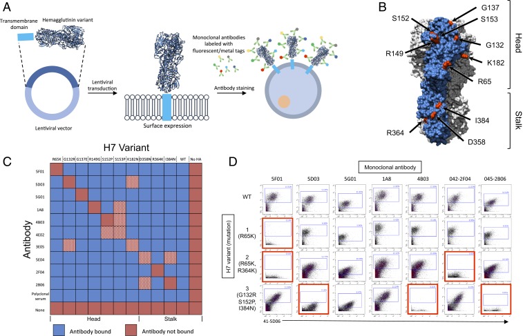
Fig. 1.
(A) Schematic of barcoding approach. (B) Modeling of A/Shanghai/1/2013 H7 HA was done using PyMOL (Protein Data Bank ID code 4LN3). Escape mutation sites highlighted in red. Single monomer highlighted in blue. (C) Heat map of monoclonal antibody binding to HEK-293T cells transfected with H7 escape mutants. Antibody–escape mutant pairs removed from the library due to cross-reactivity in white and red hatched squares. (D) Wild-type or H7 variants analyzed by mass cytometry (CyTOF). Loss of antibody binding highlighted in red.

