Fig. 6.
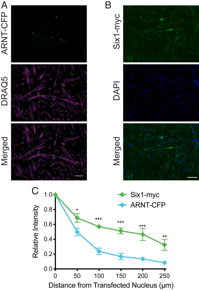
Propagation of ARNT-CFP and Six1-myc is similar to that of RFP-cNLS fusion proteins. Myoblasts were transfected with an expression vector for either aryl hydrocarbon receptor nuclear translocator fused to cyan fluorescent protein (ARNT-CFP; ∼118 kDa) or sine oculis homeobox 1 bearing a myc-tag (Six1-myc). (A and B) After 3 d of differentiation, the distributions of ARNT-CFP within DRAQ5-stained myotubes (A) or Six1-myc within DAPI-stained myotubes (B) were visualized by epifluorescence microscopy. (C) Transcription factor propagation was quantified by measuring the positions and average intensity of myonuclei within individual transfected myotubes. Distances and fluorescence intensities were normalized to the brightest myonucleus within each myotube (assumed to be the transfected nucleus). Data are binned by distance (bin size, 50 µm) and are represented as mean ± SEM (two-way ANOVA with Bonferroni posttest). (Scale bar: 100 µm.) *P < 0.05; **P < 0.01; ***P < 0.001.

