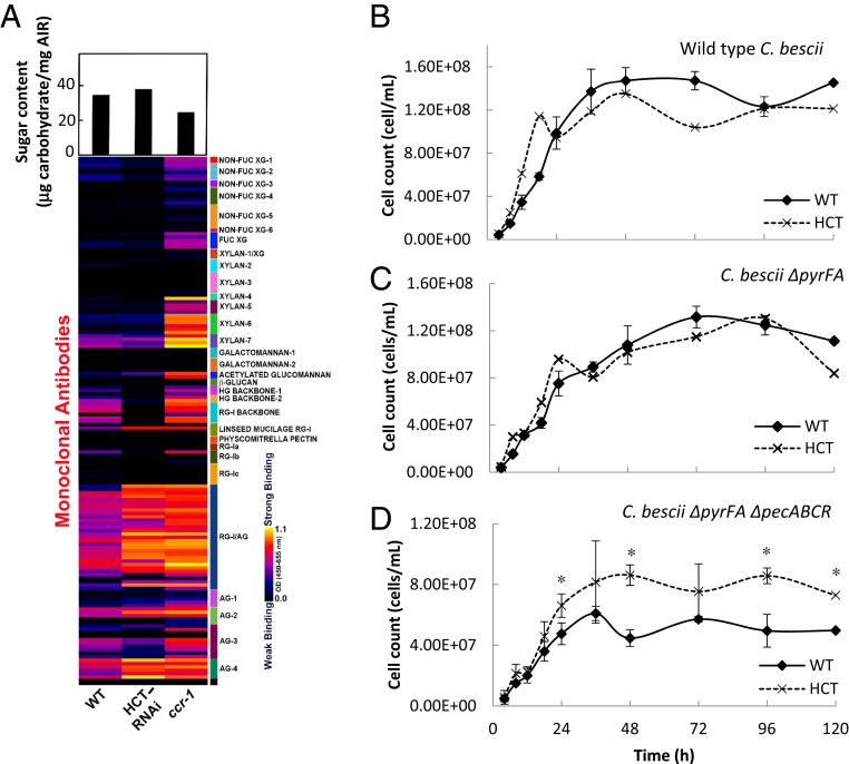
Fig. 1.
Cell wall remodeling in reduced lignin Arabidopsis lines. (A) (Lower) Glycome analysis of cell wall polysaccharides from water extracts of cell walls of control, HCT-RNAi, and ccr1 mutant Arabidopsis stems. The nondialyzed water-soluble cell wall extracts were screened against a panel of 155 monoclonal antibodies directed against diverse epitopes in noncellulosic plant glycans. The resulting heat map depicts antibody binding strength based on optical density (OD) depicted as a color gradient ranging from black (no binding) to yellow (strongest binding), as indicated by the key at the lower right. Antibodies are grouped into clades according to the glycans that they predominantly recognize, as indicated by the panel on the right side of the glycome profiles. Upper shows carbohydrate recovery from water-extracted AIRs. Details of monoclonal antibodies are given in ref. 29. (B–D) Lignin modification alleviates the need for pectinase action to enable a cellulolytic bacterium to access Arabidopsis biomass. (B) Growth of wild-type (WT) C. bescii on ground biomass from WT and HCT-down-regulated Arabidopsis plants. (C) Growth of C. bescii JWCB005 (ΔpyrFA). (D) Growth of C. bescii JWCB010 (ΔpyrFA ΔpecABCR). Cells were collected and stained with acridine orange at the times shown and counted using an epifluorescence microscope with a counting chamber lens. Two biological replicates were taken. Asterisks indicate significant differences from WT (P < 0.05) by pairwise multiple comparison Tukey test.

