Figure 2.
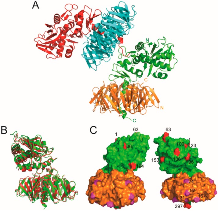
Structure of Stx2kE167Q. (A) The unit cell contained two biological units of Stx2kE167Q, each of them is constituted by an A-subunit (shown in red and green, respectively) and five B-subunits (shown in cyan and gold, respectively). The C-terminus of one of the A-subunits (shown in red) is involved in the packing of the two biological units. The N- and C-terminals of one Stx2kE167Q subunits are labeled. (B,C) Structure comparison of Stx2a and Stx2kE167Q. (B) The structure of one of the biological units of Stx2kE167Q (green) is superposed with that of Stx2a (red) and shown as a ribbon diagram. (C) A surface presentation of Stx2kE167Q with the A-subunit shown in green, and the B-subunit in gold. Residues in subunit A that are not identical with those in Stx2a is shown in red. Residues in subunit B that are not identical with those in Stx2a are shown in magenta. The non-identical residues in the A-subunit and one of the B-subunits are numbered.

