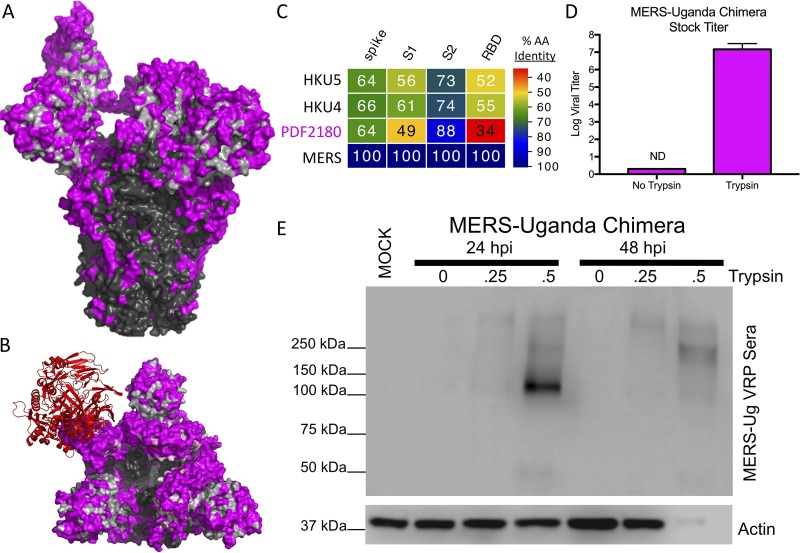
FIG 1.
Exogenous trypsin rescues MERS-Uganda spike replication. (A and B) Structure of the MERS-CoV spike trimer in complex with the receptor human DPP4 (red) from the side (A) and top (B). Consensus amino acids are outlined for the S1 (gray) and S2 (black) domains, with PDF-2180 differences noted in magenta. (C) Spike protein sequences of the indicated viruses were aligned according to the bounds of total spike, S1, S2, and receptor-binding domain (RBD). Sequence identities were extracted from the alignments, and a heatmap of sequence identity was constructed using EvolView (www.evolgenius.info/evolview) with MERS-CoV as the reference sequence. (D) MERS-Uganda chimera stocks were grown in the presence or absence of trypsin and were quantitated by plaque assay with a trypsin-containing overlay (n = 2). (E) Protein expression of MERS-Uganda spike (S) and actin 24 and 48 hours postinfection of Vero cells in the presence of increasing amounts of trypsin (none, 0.25 μg/ml, and 0.5 μg/ml) in the media.

