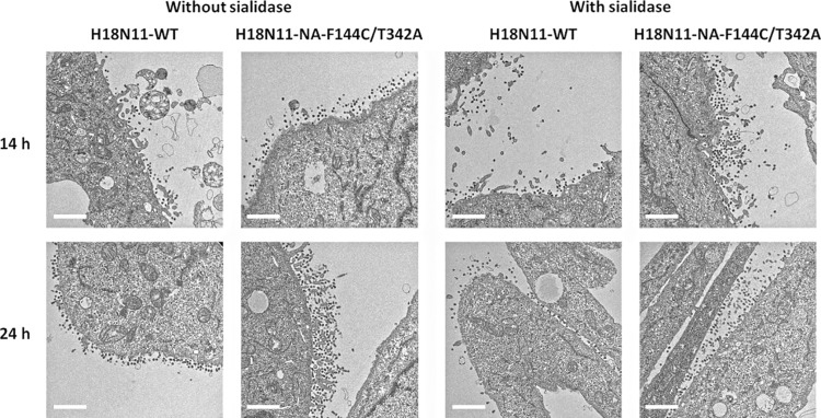
FIG 5.
Virus budding observed by use of transmission electron microscopy. MDCK II cells were infected with the H18N11-WT or H18N11-NA-F144C/T342A virus at an MOI of 5. After a 1-h incubation at 37°C, the cells were washed with PBS and Opti-MEM medium with or without 20 mU/ml sialidase was added. At 14 or 24 h postinfection, chemically fixed samples were prepared and ultrathin (110-nm-thick) sections were stained with 2% uranyl acetate and Reynold’s lead. Images were acquired with a Tecnai F20 TEM (FEI Company, Eindhoven, the Netherlands) at 200 kV. Bars, 1 μm.

