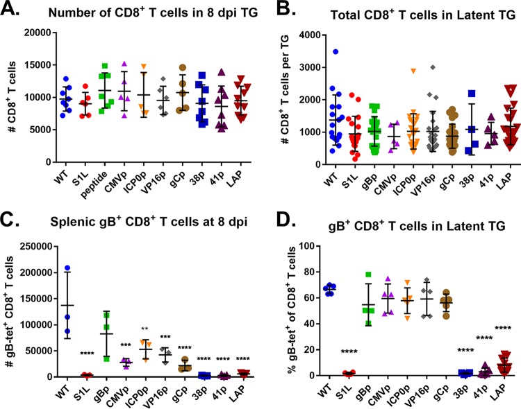
FIG 5.
gB498–505-specific CD8+ T cell responses to selected gB498–505 promoter viruses. Corneas of mice were infected with 1 × 10⁵ PFU/eye of HSV-1 WT, S1L, or a recombinant HSV-1 expressing gB498–505 from the indicated promoter. At 8 dpi (peak CD8+ T cell infiltrate) or 30 dpi (latency), TG and spleen samples were dissociated into single-cell suspensions and surface stained with antibodies to CD45, CD3, CD8, and with MHC-I gB498–505 tetramer, as detailed in Materials and Methods. (A and B) Cells were subsequently analyzed by flow cytometry and show total CD8+ T cells per TG at 8 dpi (A) or 30 dpi (B). (C) The total number of gB498–505 tetramer-positive CD8+ T cells in each spleen at 8 dpi are also shown. (D) The fraction of gB498–505 tetramer-positive cells among the total CD8+ T cells in the TG at day 30 is shown. The data shown are pooled from 2 to 3 identical experiments or are representative of one of at least two repeats of n = 3 to 5 mice per group. Bars represent the mean and standard deviation for each group. Significant differences by one-way ANOVA from the WT group are indicated *, P < 0.05; **, P < 0.01; ***, P < 0.001; ****, P < 0.0001. tet, tetramer.

