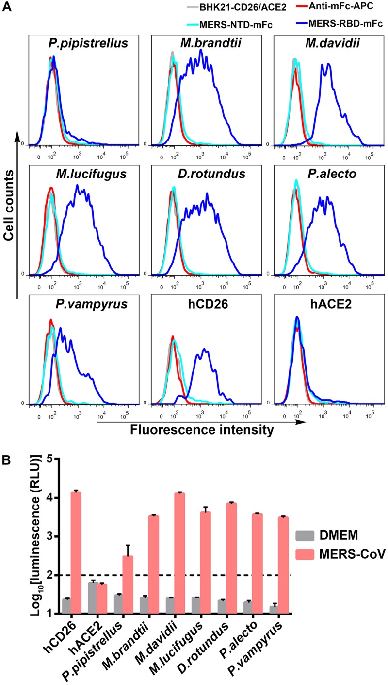
FIG 3.
Evaluation of MERS-RBD binding to bCD26s by flow cytometry and the ability of bCD26s to support the entry of pseudotyped MERS-CoV. (A) BHK21 cells transiently expressing the indicated protein, which are marked above the boxes, were stained with MERS-RBD-mFc (cyan line) or MERS-NTD-mFc (blue line). In each subplot, the gray line indicates cells without staining. The red line represents cells incubated with the secondary antibody (anti-mFc/APC). The data were collected using a BD FACSCanto and analyzed by FlowJo 7.6. The data are representative of two independent experiments. (B) Infection by lentiviral particles pseudotyped with MERS-CoV S protein. BHK21 cells, which transiently expressed the indicated proteins, were sorted based on the eGFP expression and seeded overnight. The cells were then infected with the pseudotyped MERS-CoV for 5 h. After an additional 48 h of culture, the luciferase activity was determined using a GloMax 96 microplate luminometer (Promega). DMEM without pseudotyped MERS-CoV was used as a negative control. The luminescence value for BHK21 cells with the indicated gene expression was analyzed and transformed into log scale using Prism 6. The values in each column represent the means ± the standard deviations of three replicates. The data displayed are representative of two independent experiments.

